High Content Imaging System
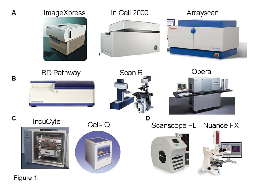
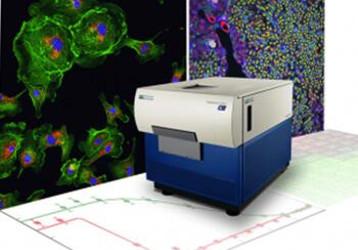
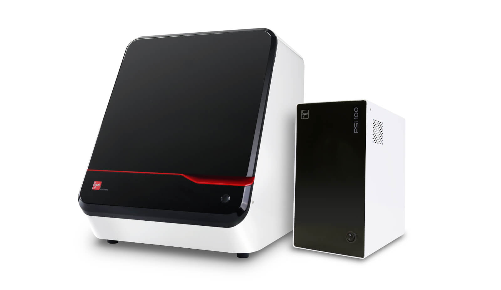
High content imaging system. Paired with our advanced analysis software our HCA systems provide high quality 3D imaging for enhanced live cell analysis. High content cell imaging and analysis Fixed and kinetic live cell assays Apoptosis Autophagy Cell cycle Cell counting Cell growthproliferation Cell migration Cell morphology Cell viabilitytoxicity Co-localization Confluency Cytotoxicity Histology Immunofluorescence NF-κB translocation Organoidspheroid morphology Transfection efficiency. The cellWoRx High Content Cell Analysis System is a wide-field imaging system from Applied Precision and Cellomics.
Our high-content confocal imaging systems based on the CSU are the ideal solution for drug screening using elaborated 3D and live cell assay systems as well as 2D fixed samples. Here are some of its key features. They feature options and modules to address your specific research including objectives filters imaging modes and.
Ultra high content imaging using MICS technology on the MACSima Imaging Platform. Its MICS MACSima TM Imaging Cyclic Staining. Our sister companies Leica Microsystems and Molecular Devices will continue to support HCA solutions.
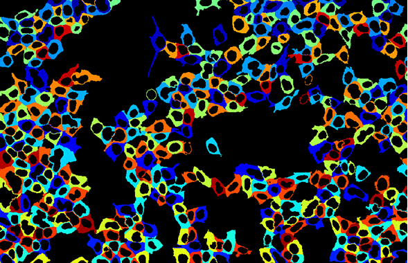
The MACSima Imaging Platform is the only complete solution for ultra high content imaging experiments. Reliable and fast cell segmentation. At its core it includes the MACSima Imaging System a fully automated instrument based on fluorescence microscopy.
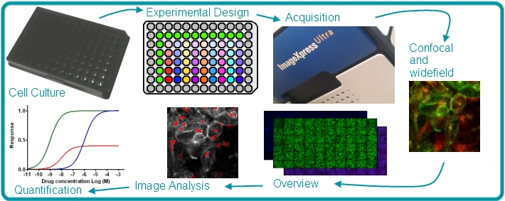
Customizable imaging protocols image-based and laser autofocusing modules and a motorized XYZ stage simplify well plate imaging and slide scanning. Yokogawas high-content analysis systems HCA also known as high-content screening systems HCS address a range of research applications from basic science to drug discovery screening. Our High-Content Screening HCS also known as High-Content Analysis HCA or High-Content Imaging platforms are designed for exceptional single-cell analysis capabilities and lightning-fast time-to-data.
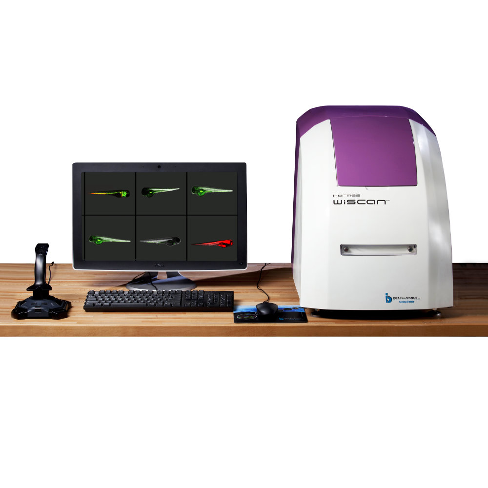
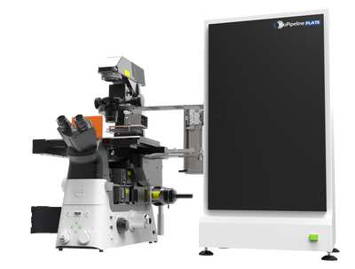
The CELENA X High Content Imaging System is an integrated imaging system designed for rapid high content image acquisition and analysis. It can capture high quality images of whole organisms thick tissues 2D and 3D models and cellular or intracellular events. WiScan Hermes IDEA Bio-Medicals automated imaging system for high content screening HCS provides the unique combination of the two contradicting primary functions of automated microscopy.
Since it is a widefield system the depth-of-field is not restricted as it is in confocal systems. Image quality and acquisition speed.
Its MICS MACSima TM Imaging Cyclic Staining.
CELENA X High Content Imaging System. CELENA X High Content Imaging System. Our High-Content Screening HCS also known as High-Content Analysis HCA or High-Content Imaging platforms are designed for exceptional single-cell analysis capabilities and lightning-fast time-to-data. Paired with our advanced analysis software our HCA systems provide high quality 3D imaging for enhanced live cell analysis. An example of a custom segmentation tool to detect colloids in. It can capture high quality images of whole organisms thick tissues 2D and 3D models and cellular or intracellular events. Reliable and fast cell segmentation. Our high-content confocal imaging systems based on the CSU are the ideal solution for drug screening using elaborated 3D and live cell assay systems as well as 2D fixed samples. The cellWoRx System provides high contrast images of samples commonly used in HCS.
Ultra high content imaging using MICS technology on the MACSima Imaging Platform. Ad Reduce artifacts by back-tracking the data to individual cellevent for quality control. High content cell imaging and analysis Fixed and kinetic live cell assays Apoptosis Autophagy Cell cycle Cell counting Cell growthproliferation Cell migration Cell morphology Cell viabilitytoxicity Co-localization Confluency Cytotoxicity Histology Immunofluorescence NF-κB translocation Organoidspheroid morphology Transfection efficiency. CELENA X High Content Imaging System. User friendly high content analysis software CellPathfinder with its versatile functionalities powerfully supports the analysis of various phenotype changes and target reactions. Paired with our advanced analysis software our HCA systems provide high quality 3D imaging for enhanced live cell analysis. NIS-Elements includes several analysis workflows as well as capabilities for user-defined custom assays that can be applied to images.











Post a Comment for "High Content Imaging System"